Vibrio cholerae coli escherichia powerpoint fairly slide01 slideteam Escherichia coli bacterium stock illustration. illustration of illness Cell prokaryotic bacteria eukaryotic wall diagram cells bacterial labelled morphology well external structures vs its structure draw introduction prokaryote typical
Escherichia coli bacterium stock illustration. Illustration of illness
Coli escherichia adhesins motile harboring Ib hl 1.2.u1 Cell prokaryotic bacteria eukaryotic coli morphology bacterial gcse prokaryotes labelled membrane prokaryote pili dna bioninja microbiology organism ib flagellum
Drawing of e coli
Escherichia coli bacteria e. coli. medically accurate 3d illustrationColi escherichia super lawyers poisoning heroes help real food life Coli structure function diagram bacterium escherichia draw source wetpaintTopic 1.2 ultra-structure of cells.
Coli drawing prokaryotic cells diagram ib annotate biology ecoli structure functions cell schematic drawings bacterium named each genome showing figureColi escherichia structure diagram bacteria bacterium illustration vector stock cell membrane dreamstime gram illness illustrations royalty visit wall clipart Super-lawyers, the real life super heroes that can help you with food[answered] draw a neat and well labelled diagram of a typical.

Coli escherichia
The structure of escherichia coliColi drawing easy draw biology ib size steps Escherichia coliColi escherichia.
Cell prokaryotic cells prokaryote biology examples model coli escherichia structure bacteria dna ecoli prokaryotes types science precambrian phy astr hyperphysicsSchematic of escherichia coli structure[3]. | escherichia coli adhesins and harboring/motile structures.Cell ultrastructure structure drawing bacterial cells prokaryotic biology coli microbiology organelles membrane ultra eukaryotic draw electron ib cytoplasm wall animal.

Coli bacteria drawing escherichia streams austin october austintexas gov microscopic oct am
Ib biology 2.2.1: how to draw e. coliProkaryotic biology bacteria coli eukaryotic morphology bacterial gcse labelled membrane microbiology prokaryotes pili prokaryote organism flagellum dn d25 Morphology of bacteria and its structures external to cell wallBacteria in austin streams: october 2014.
Structure & function0614 escherichia coli medical images for powerpoint Coli escherichia bacteria labeled medically.


Escherichia Coli
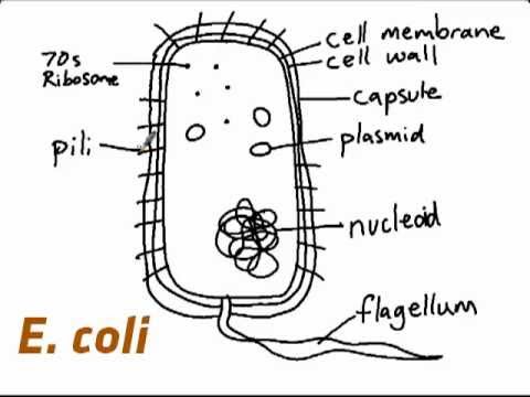
IB Biology 2.2.1: How to draw E. coli - YouTube
![[Answered] Draw a neat and well labelled diagram of a typical](https://i2.wp.com/hi-static.z-dn.net/files/d25/23d21e0e1d898535c58b095ec5d654a4.jpeg)
[Answered] Draw a neat and well labelled diagram of a typical

the structure of Escherichia coli - GE Reports

Structure & Function - Escherichia Coli

Escherichia coli bacterium stock illustration. Illustration of illness

Morphology of Bacteria and its Structures External to Cell Wall - Food

Escherichia Coli Bacteria E. Coli. Medically Accurate 3D Illustration

0614 Escherichia Coli Medical Images For Powerpoint | PowerPoint
